
What Is a DEXA Scan — and How Is It Used in Assessing Bone Health?
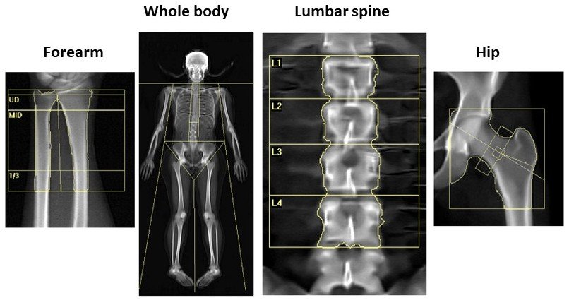
A DEXA scan is the investigation most commonly used to measure bone density and help assess the risk of osteoporosis and future fractures.
People may be referred for a scan after a fragility fracture, because they take long-term steroids, have experienced an early menopause or have a medical condition that can affect bone health. Others receive a report containing terms such as “osteopenia”, “T-score” or “low bone density” without a clear explanation of what those findings mean for them.
A DEXA result is important, but it is not a complete assessment on its own. Bone density must be considered alongside age, previous fractures, medication, other health conditions and the overall likelihood of sustaining a fracture.
What is a DEXA scan?
DEXA stands for dual-energy X-ray absorptiometry. Technically, the abbreviation DXA is increasingly preferred, although DEXA remains the term most patients encounter.
The scan uses two very low-energy X-ray beams to measure bone mineral density. The lumbar spine and hips are usually examined because these are clinically important sites for osteoporotic fractures. The forearm may be assessed in selected circumstances, particularly when the spine or hips cannot be interpreted reliably.
DEXA uses a very low dose of radiation. It is painless, non-invasive and does not require an injection.
What happens during the scan?
During the examination, you lie on an open scanning table while a scanning arm moves over the relevant part of the body. It is not an enclosed scanner or tunnel.
The examination usually takes approximately 10 to 20 minutes. You may be asked to remove clothing or objects containing metal, and positioning devices may be used to place the hips and spine correctly. Accurate positioning matters because small technical differences can affect the measurements.
Preparation is generally minimal, although you should follow the instructions provided by the scanning department. You should also tell the department if there is any possibility that you may be pregnant or if you have recently had an investigation involving contrast material.
Who may need a DEXA scan?
A DEXA scan is not a general investigation for joint pain, back pain or fatigue. It is most useful when the result is likely to influence an assessment of fracture risk or a decision about treatment.
A scan may be considered after a fracture caused by relatively minor trauma, with long-term or repeated glucocorticoid treatment, following premature menopause, or in people with low body weight, a strong family history of hip fracture or a medical condition associated with bone loss. Rheumatoid arthritis, inflammatory bowel disease, coeliac disease, endocrine disorders and some cancer treatments can all affect bone health.
Updated NICE guidance also allows some people with a previous hip or vertebral fragility fracture, or two or more fragility fractures, to proceed directly to DEXA without first completing a fracture-risk calculation.
In other situations, a tool such as FRAX may be used first to decide whether measuring bone density is likely to add useful information.
Understanding T-scores
A T-score compares your bone density with the average peak bone density of a healthy young adult.
T-scores are principally used to interpret results in postmenopausal women and men aged 50 or over.
T-score General interpretation
–1.0 or above Bone density within the expected young-adult range
Below –1.0 but above –2.5 Low bone mass, often described as osteopenia
–2.5 or below Bone density in the osteoporosis range
These categories are useful, but they should not be interpreted as automatic treatment instructions.
Osteopenia is a description of bone density, not a disease requiring medication in every case. Some people with osteopenia have a high fracture risk because of their age, fracture history or other risk factors. Others have a relatively low risk and may not need medication.
Similarly, a T-score in the osteoporosis range is important, but the treatment decision still needs to account for fracture history, clinical circumstances and whether the measurement is technically reliable.
What is a Z-score?
A Z-score compares your bone density with that of people of a similar age and sex.
Z-scores are generally preferred in premenopausal women and men under 50. A Z-score of –2.0 or lower is described as being “below the expected range for age”.
This does not, by itself, diagnose osteoporosis in a younger person. Instead, it may prompt consideration of why bone density is lower than expected. Possible contributors include hormonal conditions, malabsorption, inflammatory disease, medication, low body weight or another underlying medical problem.
This distinction is important because the familiar T-score thresholds should not be applied in exactly the same way to every age group.
What can a DEXA scan tell us?
DEXA provides a reliable measurement of bone mineral density. It can help identify low bone mass or osteoporosis, contribute to fracture-risk assessment and provide a baseline against which future measurements may be compared.
However, bone density is only one component of bone strength. A standard DEXA scan does not directly measure the microscopic structure or quality of bone. It does not explain why bone loss has occurred, and it cannot predict precisely whether or when an individual will sustain a fracture.
It is also not designed to diagnose the cause of back pain, osteoarthritis or cancer.
Some centres provide a measurement called trabecular bone score, or TBS, which is calculated from the lumbar-spine image and can add information about bone texture. It may be incorporated into fracture-risk assessment in selected patients, but it is not available or required in every case.
Why might the hip and spine results be different?
It is common for the hip and spine to produce different scores. Bone loss does not necessarily occur at the same rate throughout the skeleton.
The lumbar-spine result can also be affected by osteoarthritis, degenerative change, previous vertebral fractures or calcification in nearby structures. These can make the measured spine density appear higher than it truly is.
Conversely, an individual vertebra may sometimes produce an unexpectedly low or inconsistent result because of a structural abnormality or technical issue.
Good interpretation therefore involves more than selecting the lowest number on the report. The clinician reporting the scan should consider image quality, positioning, the pattern across the measured sites and whether any area should be excluded from interpretation.
How does FRAX fit in?
FRAX estimates the 10-year probability of a major osteoporotic fracture and of a hip fracture specifically.
A major osteoporotic fracture in FRAX refers to a clinical fracture of the spine, hip, forearm or proximal humerus. The calculation considers age, sex, height, weight, previous fractures, parental hip fracture, smoking, alcohol intake, glucocorticoid use, rheumatoid arthritis and causes of secondary osteoporosis.
FRAX can be calculated without a DEXA result. When bone density is added, FRAX uses the measurement from the femoral neck—not the total hip or lumbar spine.
This is one reason FRAX and DEXA should be seen as complementary rather than competing tests. DEXA measures bone density at particular sites, while FRAX brings that information together with clinical factors that also influence fracture risk.
FRAX does not make a treatment decision by itself. It may underestimate risk in some circumstances, including multiple previous fractures, vertebral fractures, high-dose steroid treatment and conditions associated with particularly rapid bone loss. Clinical judgement remains essential.
Can a DEXA scan detect spinal fractures?
A standard DEXA scan measures bone density and is not primarily a fracture-detection test. However, some DEXA machines can perform a vertebral fracture assessment, usually abbreviated to VFA.
VFA is a low-dose image of the spine that can identify vertebral fractures which may otherwise go unrecognised. These fractures do not always cause sudden severe pain. They may instead present with height loss, increasing spinal curvature or persistent unexplained back discomfort.
Current NICE guidance recommends considering VFA in a wider group of people undergoing DEXA, depending on age and additional risk factors. Finding a vertebral fracture can materially change the assessment because a previous fragility fracture is a strong predictor of future fractures.
VFA is not required for everyone, and conventional spinal imaging may still be necessary when symptoms or findings need more detailed assessment.
Why might blood tests be needed?
DEXA measures bone density but does not identify the cause of bone loss.
Depending on the circumstances, blood tests may be used to look for vitamin D deficiency, kidney disease, thyroid or parathyroid disorders, coeliac disease, abnormalities of calcium or phosphate, and other conditions that can affect bone metabolism.
The investigations should be guided by the clinical history rather than ordered as the same large panel for everyone.
What are bone-turnover markers?
Bone is constantly being broken down and rebuilt. Blood tests known as bone-turnover markers can provide information about these processes.
CTX reflects bone resorption, while P1NP reflects bone formation. These tests are not routinely needed to diagnose osteoporosis and they do not replace DEXA or fracture-risk assessment.
Their main value is in selected situations, such as checking biological response or adherence to treatment and helping guide the sequencing of certain osteoporosis therapies. Results require careful interpretation because CTX in particular can vary with food intake, time of day, kidney function and recent fractures.
How often should DEXA be repeated?
Bone density usually changes slowly. Repeating a scan too soon may show differences that are smaller than the expected measurement variation and therefore difficult to interpret reliably.
Most people do not need another DEXA scan within two years. An interval of approximately two to five years is common, but the timing should be individualised according to the initial result, age, treatment, fracture history and any medical condition associated with rapid bone loss.
Shorter intervals may occasionally be appropriate with glucocorticoid treatment, some cancer therapies, anabolic osteoporosis treatment or another situation in which bone density may change more quickly.
Comparison is most reliable when the scans are performed using the same machine or properly cross-calibrated equipment. A small numerical difference does not necessarily represent genuine bone loss or gain. The change must exceed the facility’s calculated “least significant change” before it can confidently be regarded as real.
Does an abnormal DEXA result automatically mean treatment?
No. Treatment is intended to reduce fracture risk rather than simply improve a number on a scan.
Some people with osteopenia need medication because their overall fracture risk is high. Others with a similar T-score can reasonably be monitored without treatment. Conversely, a person who has sustained a hip or vertebral fragility fracture may require treatment even when the DEXA result is not in the osteoporosis range.
Management may involve resistance and weight-bearing exercise, adequate protein and nutrition, correction of vitamin D or calcium deficiency, reducing falls risk and addressing smoking or excessive alcohol intake.
When medication is appropriate, the choice depends on the degree of fracture risk, previous treatment, kidney function, other medical conditions and the need for a safe long-term sequence. Options may include anti-resorptive treatment or, for selected people at very high fracture risk, bone-building treatment.
When can specialist interpretation be helpful?
Many DEXA reports can be interpreted and managed appropriately in primary care or through a fracture liaison service. Specialist assessment may be helpful when the findings are inconsistent, when fractures have occurred despite apparently reassuring bone density or when there is uncertainty about the underlying cause.
It may also be useful in younger people with unexpectedly low bone density, people with complex secondary osteoporosis, those who cannot tolerate standard treatment and patients being considered for injectable or bone-building therapies.
The purpose of specialist assessment is not simply to prescribe medication. It is to determine whether treatment is needed, explain the available options and ensure that any plan is safe and sustainable over the longer term.
If you have an abnormal DEXA result, a previous fragility fracture or uncertainty about whether treatment is needed, specialist assessment can place the scan findings in the context of your overall fracture risk. Information about private bone-health consultations is available here.
The bottom line
A DEXA scan measures bone mineral density, usually at the hip and lumbar spine. In postmenopausal women and men aged 50 or over, results are generally interpreted using T-scores. In younger adults, Z-scores are usually more appropriate.
The scan provides valuable information, but it does not measure every aspect of bone strength and should not be interpreted in isolation. Previous fractures, age, medication, falls risk and underlying medical conditions may be just as important as the number on the report.
FRAX, vertebral fracture assessment, selected blood tests and occasionally bone-turnover markers can add further information. The aim is not simply to categorise a scan as normal, osteopenic or osteoporotic. It is to understand the individual’s actual fracture risk and make a proportionate long-term plan.
Related reading
Understanding Bone Health and Osteoporosis: What Patients Should Know
Bisphosphonates for Osteoporosis: Benefits, Side Effects and How Long to Take Them
Denosumab for Osteoporosis: What It Does and Why Stopping Needs a Plan
Romosozumab in Osteoporosis: When and Why It Is Used
Trusted patient information
Royal Osteoporosis Society: Bone density scans
Please note, these posts are for general information only and do not constitute medical advice. Dr Singh would encourage you to speak to your healthcare professional to be assessed and managed for your specific symptoms.